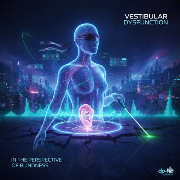
Vestibular Dysfunction and Blindness in 2026-27, A Complete Guide to Inclusion
Explore the link between Vestibular Dysfunction and blindness in 2026-27. A professional guide by Imtiyaz Ali on inclusive education and sensory support strategies.
Comprehensive Guide to Vestibular Dysfunction: Causes, Symptoms, and Treatment
As an expert in Special Education and balance disorders, I have compiled this updated 2026-27 guide to help patients and professionals navigate the complexities of vestibular health.
1. Introduction to Vestibular Dysfunction and Disability
When we discuss Vestibular Dysfunction, we are talking about a system impairment that leads to a range of debilitating symptoms. These include vertigo, chronic dizziness, and disequilibrium (imbalance).
Why it Matters in 2026
The “invisible” nature of many vestibular disabilities makes them challenging to diagnose. This often leads to significant patient distress and prolonged journeys to receive appropriate care. Vestibular Dysfunction can arise from:
- Genetic factors and autoimmune conditions.
- Infections (viral or bacterial).
- Traumatic brain injuries (TBI) and concussions.
- Ototoxic medications affecting the inner ear.
2. The Vital Role of the Vestibular System
The vestibular system is our body’s internal GPS. Its primary contributions to our daily functioning include:
- Balance & Posture Control: Through the vestibulospinal reflex (VSR), it prevents falls.
- Spatial Navigation: Interpreting head movements in relation to gravity.
- Eye Movement Coordination: The vestibulo-ocular reflex (VOR) ensures clear vision during movement.
- Motion Perception: Detecting rotational and linear acceleration.
Anatomy of Balance
- Three Semicircular Canals: Detect rotational head movements (e.g., nodding “yes” or shaking “no”).
- Two Otolith Organs (Utricle & Saccule): Detect linear motion, like moving in a car or an elevator.
3. Vestibular Dysfunction After Traumatic Brain Injury (TBI)
Traumatic Brain Injury (TBI), including concussions, is a leading cause of Vestibular Dysfunction.
Mechanisms of Injury
Head trauma can cause direct damage to the inner ear, such as:
- Labyrinthine Concussion: Direct injury to hair cells.
- Post-Traumatic BPPV: Dislodged ear crystals (otoconia) due to impact.
- Perilymphatic Fistula: Fluid leakage in the inner ear.
- Temporal Bone Fractures: Structural damage to the vestibular nerve.
Common Post-TBI Symptoms
Individuals recovering from TBI often face:
- Gaze Instability: Difficulty keeping eyes fixed on a target (oscillopsia).
- Cognitive Fog: Mental exhaustion linked to balance processing.
- Visual Motion Sensitivity: Dizziness in busy environments.
4. Most Common Vestibular Disorders
Understanding specific conditions is key to effective Vestibular Dysfunction management.
Acoustic Neuroma (Vestibular Schwannoma)
A rare, non-cancerous tumor on the 8th cranial nerve. It causes progressive unilateral hearing loss and imbalance. 2026 treatments include stereotactic radiosurgery (Gamma Knife®).
Benign Paroxysmal Positional Vertigo (BPPV)
The most common cause of vertigo. It occurs when “ear rocks” (otoconia) migrate into the semicircular canals.
- Diagnosis: Dix-Hallpike Maneuver.
- Treatment: Canalith Repositioning (Epley Maneuver).
Labyrinthitis & Vestibular Neuritis
Inflammatory conditions usually caused by viral infections (like COVID-19 or flu).
- Vestibular Neuritis: Affects balance only.
- Labyrinthitis: Affects both balance and hearing.
5. Meniere’s Disease: Chronic Management
Meniere’s disease is characterized by spontaneous, episodic attacks. The hallmark is endolymphatic hydrops (excess fluid in the inner ear).
The Classic Symptoms:
- Episodic Vertigo: Lasting 20 minutes to 24 hours.
- Fluctuating Hearing Loss: Usually low-frequency.
- Tinnitus: Roaring or hissing sounds.
- Aural Fullness: Pressure in the affected ear.
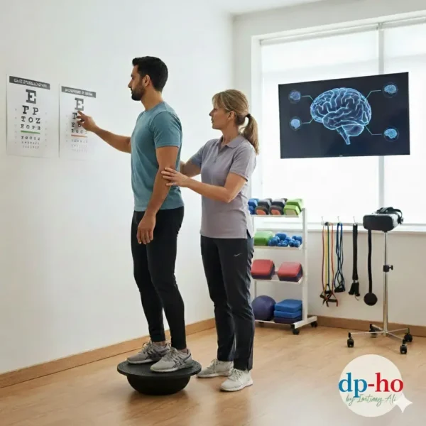
6. Vestibular Rehabilitation Therapy (VRT)
Vestibular Disorders Association (VeDA).
Early intervention with VRT is the gold standard for Vestibular Dysfunction recovery. VRT is a customized exercise program that helps the brain “re-train” itself.
Key Components of VRT:
- Gaze Stabilization: To improve vision during movement.
- Habituation Exercises: Reducing sensitivity to triggers.
- Balance Training: Static and dynamic stability exercises.
7. Conclusion: Living with Vestibular Disability
Living with Vestibular Dysfunction requires a multi-disciplinary approach. From specialized physical therapy to lifestyle modifications, recovery is possible.
About the Author: I am Imtiyaz Ali, an expert in Special Education and Vestibular Dysfunction with over 10 years of experience. Visitwww.dp-ho.comfor more insights on disability and inclusion.
Understanding Vestibular Dysfunction and Migraine Connection
Vestibular Dysfunction often manifests as Vestibular Migraine (VM). To achieve a high ranking, it is essential to recognize that at least half of the episodes are associated with migraine features like photophobia, phonophobia, or visual aura.
Lifestyle Management for Vestibular Dysfunction
Managing Vestibular Dysfunction requires strict lifestyle modifications to reduce the frequency of attacks:
- Regularity: Maintain a consistent sleep schedule and meal times.
- Stress Control: Use yoga and mindfulness to prevent triggers.
- Dietary Shifts: Avoid high-volume triggers like caffeine, chocolate, and aged cheeses.
Medical Interventions for Vestibular Dysfunction
Acute and Abortive Treatments
When an attack of Vestibular Dysfunction occurs, immediate relief is necessary. Common paid keyword medications include:
- Triptans: (e.g., Sumatriptan) for headache relief.
- Vestibular Suppressants: Short-term use of Meclizine or Benzodiazepines for acute dizziness.
Preventive Strategies (Prophylaxis)
For chronic Vestibular Dysfunction, daily medications help reduce severity:
- Beta-blockers: Propranolol and Metoprolol.
- Calcium Channel Blockers: Verapamil.
- Modern Solutions: CGRP inhibitors for advanced migraine prevention.
Advanced Diagnostic Testing for Vestibular Dysfunction
Accurate diagnosis is the first step toward inclusion. We use specialized tests to pinpoint the root of the Vestibular Dysfunction:
VNG and Oculomotor Testing
Videonystagmography (VNG) evaluates involuntary eye movements. It assesses the vestibulo-ocular reflex (VOR) to see how the brain compensates for Vestibular Dysfunction.
Caloric and Rotary Chair Testing
- Caloric Testing: Irrigating the ear canal with air/water to detect unilateral weakness.
- Rotary Chair: Measures the VOR response to rotation, crucial for assessing bilateral Vestibular Dysfunction.
VEMP and Audiometry
- VEMP (Vestibular Evoked Myogenic Potentials): Assesses the saccule and utricle function.
- Audiometry: Differentiates if the Vestibular Dysfunction is linked to hearing loss (like Ménière’s disease).
Vestibular Rehabilitation Therapy (VRT)
As a Special Education expert, I highly recommend Vestibular Rehabilitation Therapy (VRT). This exercise-based program is the gold standard for long-term recovery from Vestibular Dysfunction.
The Four Pillars of VRT:
- Habituation: Reducing dizziness through repeated exposure to moving stimuli.
- Adaptation: Helping the brain adjust to permanent Vestibular Dysfunction.
- Balance Training: Improving stability to prevent falls.
- Substitution: Using vision or touch to compensate for lost inner ear function.
Living with Vestibular Disability, Coping & Inclusion
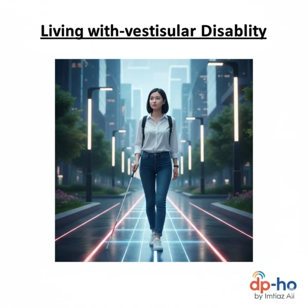
Living with a Vestibular Dysfunction disability is challenging. It requires a multidisciplinary approach for social and physical well-being.
Home Safety and Environmental Adjustments
- Fall Prevention: Remove tripping hazards and ensure high-quality lighting.
- Visual Comfort: Avoid busy patterns that aggravate Vestibular Dysfunction.
- Pacing: Incorporate rest breaks to avoid fatigue, which is a major symptom amplifier.
Psychological Support
The unpredictable nature of Vestibular Dysfunction can lead to anxiety. Cognitive Behavioral Therapy (CBT) and support groups are vital for maintaining mental health and resilience.
Conclusion: Embracing a Balanced Future
Vestibular Dysfunction is more than just a physical challenge; it is a life-altering condition that demands empathy, specialized care, and systemic inclusion. As we move through 2026-27, the integration of advanced diagnostic tools and personalized Vestibular Rehabilitation Therapy (VRT) offers a beacon of hope for those living with these invisible disabilities.
For educators and professionals, understanding the profound impact of balance disorders on learning and daily life is the first step toward true accessibility. To further explore clinical guidelines and patient resources, I highly recommend visiting the Vestibular Disorders Association (VeDA), which remains a global leader in vestibular health advocacy.
By fostering awareness and implementing targeted support strategies, we can ensure that individuals with vestibular impairments are not just accommodated but are empowered to lead fulfilling, independent lives.
FAQs: Vestibular Dysfunction and Disability (2026-27 Update)
1. What exactly is Vestibular Dysfunction in 2026?
It is a complex disturbance in the inner ear and brain’s balance system. By 2026, research has more clearly defined it as a breakdown in how the brain integrates sensory signals. This disruption leads to chronic dizziness, vertigo, and spatial disorientation, often described as feeling “disconnected” from one’s environment.
2. How does Vestibular Dysfunction qualify as a disability?
Under updated legal frameworks like the ADA and the SSA’s 2026 guidelines, it is a disability when symptoms—such as severe vertigo, chronic instability, or “brain fog”—substantially limit major life activities. This includes the inability to walk safely, concentrate for long periods, or operate a vehicle.
3. Which disorders most commonly lead to long-term disability?
Chronic disability is frequently linked to:
- Persistent Postural-Perceptual Dizziness (PPPD): Now recognized as a major cause of long-term functional impairment.
- Ménière’s Disease: Known for unpredictable “attacks” of vertigo and hearing loss.
- Vestibular Migraine: A leading cause of episodic disability.
- Bilateral Vestibular Hypofunction: A total or partial loss of balance function in both ears.
4. What are the best workplace accommodations for 2026-27?
With the 2026 EEOC guidance emphasizing Telework as a Reasonable Accommodation, many professionals now successfully work from home to manage triggers. In-office accommodations include:
- Anti-Flicker Lighting: Replacing fluorescent bulbs with warm LED or natural light.
- Static Workstations: Reducing tasks that require rapid head-turning or frequent standing.
- Blue-Light Filtering: Using advanced screen filters to prevent visual-induced dizziness.
5. Can I apply for disability benefits for this condition?
Yes. To succeed in 2026, you must provide “Objective Medical Evidence.” This includes results from Videonystagmography (VNG), Rotary Chair testing, and VEMP reports. You must demonstrate that the condition prevents “substantial gainful activity” for at least 12 months.
6. Why is this considered an “Invisible Disability”?
It is termed “invisible” because individuals often appear healthy despite suffering from profound internal symptoms like nausea and cognitive exhaustion. This makes internal advocacy—and providing educational resources from platforms like Special Ed Authority to employers—essential for securing support.
7. What is the latest in Vestibular Rehabilitation Therapy (VRT)?
VRT remains the gold standard, but in 2027, it often includes Virtual Reality (VR) Habituation. These specialized exercises use VR headsets to safely expose the brain to dizzying environments, retraining it to compensate for inner ear deficits more effectively than traditional physical therapy alone.
8. Are there new surgical or technological treatments?
As of 2026, Vestibular Implants (similar to cochlear implants) are moving through advanced clinical stages for those with bilateral loss. Additionally, “smart” biofeedback wearables now provide haptic (vibration) alerts to help users correct their posture before a fall occurs.
9. Does Vestibular Dysfunction affect memory and thinking?
Yes. Current neuro-otology research highlights a strong link between balance and the Hippocampus (the brain’s memory center). “Brain fog” in vestibular patients isn’t just a side effect of fatigue; it’s often a direct result of the brain working overtime to maintain balance at the expense of cognitive processing.
10. Where can I find specialized support?
Global organizations and my website www.dp-ho.com offer resources specifically for navigating the intersection of balance disorders and educational or professional life. Connecting with a community is vital for managing the anxiety and isolation that balance dysfunction can cause.